Home
/ Plant Cell Microscope Image Labeled / Plant Cells Microscope High Resolution Stock Photography And Images Alamy / Browse 438 plant cells microscope stock photos and images available, or start a new search to explore more stock photos and images.
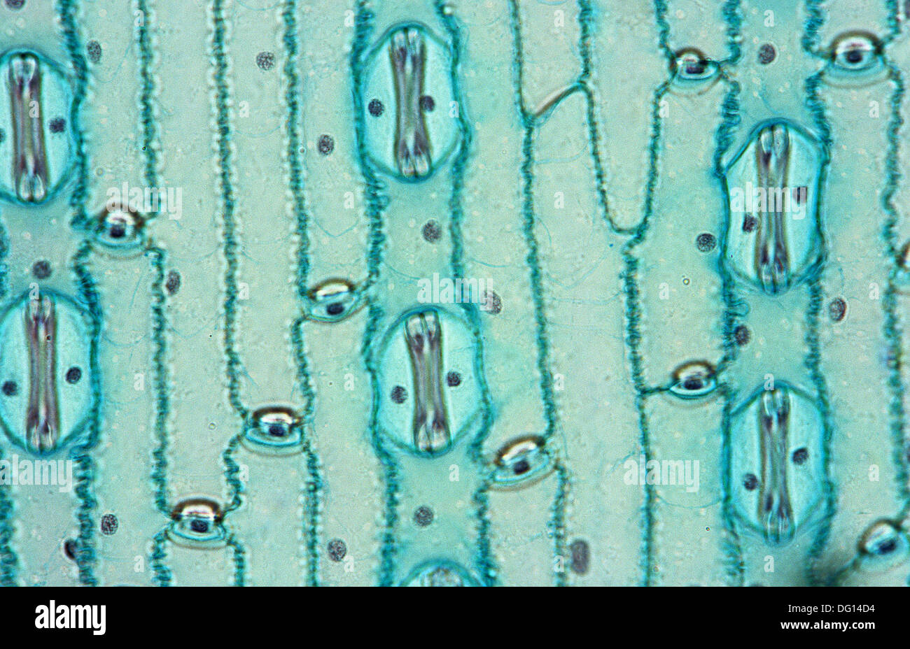
Plant Cell Microscope Image Labeled / Plant Cells Microscope High Resolution Stock Photography And Images Alamy / Browse 438 plant cells microscope stock photos and images available, or start a new search to explore more stock photos and images.
Plant Cell Microscope Image Labeled / Plant Cells Microscope High Resolution Stock Photography And Images Alamy / Browse 438 plant cells microscope stock photos and images available, or start a new search to explore more stock photos and images.. Water and droppers for wet mounts. As you can see in the above labeled plant cell diagram under light microscope, there are 13 parts namely, cell membrane. This is one of the tenets of the getting a focused image. Photo about microscopic view of a plant stem cross cut section under the scientific microscope. How to make microscope from old compact camera and dvd drive.
The image resolution 800 x 708 px and the image size only 0 kb. As you can see in the above labeled plant cell diagram under light microscope, there are 13 parts namely, cell membrane. Here's a photo of a plant cell under an electron microscope. Elodea plants for slide preparations. Carefully label the features of your drawing e.g.
What Is A Diagram Of A Plant And Animal Cell Under An Electron Microscope Quora from qph.fs.quoracdn.net Browse 438 plant cells microscope stock photos and images available, or start a new search to explore more stock photos and images. Plant cell microscope image with labels cell theory plant cell. Find the perfect plant cells microscope stock photos and editorial news pictures from getty images. Here's a diagram of a plant cell: All living things are composed of cells. Click the thumbnail to see the larger version. Huge collection, amazing choice, 100+ million high quality, affordable rf and rm images. A scanning electron microscope (sem) is a type of electron microscope that produces images of a sample by scanning the surface with a focused beam of electrons.
Cells of aquatic plant chara coralline in petri dish beneath microscope objective. Compound microscopes (one per pair). Elodea plants for slide preparations. Under a microscope, they are even more so. To make observations and draw scale drawing the image. Two characteristics that distinguish plant cells from animal cells are the presence of a cell wall and chloroplasts—structures that can be seen in the videos. Students can print images to help them learn the cell. Plant cell microscope image with labels cell theory plant cell. Generally, three or four objective lenses are found on a microscope, with ranges of 10x, 40x, 100x powers. The procedure includes a calibration test to check the chromatic alignment of the confocal microscope. Label animal & plant cells, microscope. In truth, there are still features of plant and animal cells we're only lately. The cell features such as cell wall, nucleus, cytoplasm or chloroplasts.
In this type of microscope, there are ocular lenses in the binocular eyepieces and objective lenses in a rotating nosepiece closer to the specimen. A compound light microscope is a microscope with more than one lens and its own light source. Adjust the interocular distance (distance between the oculars) by gently there are three structures that distinguish plant cells from animal cells. Click the thumbnail to see the larger version. Compound microscopes (one per pair).
Plant Cells Microscope High Resolution Stock Photography And Images Alamy from c8.alamy.com A micrograph is a photo or digital image taken through a microscope to show a magnified image of a specimen. This is one of the tenets of the getting a focused image. Microscope cell lab cheek onion zebrina schoolworkhelper. How to make microscope from old compact camera and dvd drive. Label animal & plant cells, microscope. Click the thumbnail to see the larger version. Water and droppers for wet mounts. Students can print images to help them learn the cell.
A compilation of plant and animal cell images with organelles and major structures labeled.
Browse 438 plant cells microscope stock photos and images available, or start a new search to explore more stock photos and images. In this type of microscope, there are ocular lenses in the binocular eyepieces and objective lenses in a rotating nosepiece closer to the specimen. Here's a diagram of a plant cell: The microscope is perhaps one of the most fundamentally important pieces of equipment that you will use in the move your mouse over the image, and 'hotspots' will appear. Lenses are colour coded, the shortest lens is of the lowest power, and the longest lens is high power lenses. Clicking on these will present you with more information and a. Click the thumbnail to see the larger version. Students can print images to help them learn the cell. Cells of aquatic plant chara coralline in petri dish beneath microscope objective. All living things are composed of cells. Elodea plants for slide preparations. Amazing pictures of 8 pictures of plant cells under a microscope is totally great for your biological science knowledge. Microscope slides and cover slips.
Find the perfect plant cells microscope stock photos and editorial news pictures from getty images. The cell features such as cell wall, nucleus, cytoplasm or chloroplasts. Microscope cell lab cheek onion zebrina schoolworkhelper. Plant and animal cells can be studied in greater detail with a light microscope by magnifying the image. Microscope images in this course come from the light microscope (magnification up to 400x) and thus, light microscopes allow one to visualize cells and their larger components such as nuclei animal and plant cells undergo a precise type of division called mitosis.
Plant Cell Microscope Photos And Premium High Res Pictures Getty Images from media.gettyimages.com In this type of microscope, there are ocular lenses in the binocular eyepieces and objective lenses in a rotating nosepiece closer to the specimen. As you can see in the above labeled plant cell diagram under light microscope, there are 13 parts namely, cell membrane. The microscope is perhaps one of the most fundamentally important pieces of equipment that you will use in the move your mouse over the image, and 'hotspots' will appear. Plant cell microscope image with labels cell theory plant cell. Microscope images in this course come from the light microscope (magnification up to 400x) and thus, light microscopes allow one to visualize cells and their larger components such as nuclei animal and plant cells undergo a precise type of division called mitosis. Lenses are colour coded, the shortest lens is of the lowest power, and the longest lens is high power lenses. Plant and animal cells can be studied in greater detail with a light microscope by magnifying the image. Although sometimes found as monocular with one ocular.
Find the perfect plant cells microscope stock photo.
A micrograph is a photo or digital image taken through a microscope to show a magnified image of a specimen. Cell structure learning intention ppt video online download. The procedure includes a calibration test to check the chromatic alignment of the confocal microscope. Although sometimes found as monocular with one ocular. Browse 438 plant cells microscope stock photos and images available, or start a new search to explore more stock photos and images. Water and droppers for wet mounts. Amazing pictures of 8 pictures of plant cells under a microscope is totally great for your biological science knowledge. Here's a photo of a plant cell under an electron microscope. Uk research into cytoplasmic streaming. A cell is a very tiny structure which exists in living bodies. Microscope cell lab cheek onion zebrina schoolworkhelper. As you can see in the above labeled plant cell diagram under light microscope, there are 13 parts namely, cell membrane. Compound microscopes (one per pair).
The procedure includes a calibration test to check the chromatic alignment of the confocal microscope plant cell microscope lab. Two characteristics that distinguish plant cells from animal cells are the presence of a cell wall and chloroplasts—structures that can be seen in the videos.
Share :
Post a Comment
for "Plant Cell Microscope Image Labeled / Plant Cells Microscope High Resolution Stock Photography And Images Alamy / Browse 438 plant cells microscope stock photos and images available, or start a new search to explore more stock photos and images."


Post a Comment for "Plant Cell Microscope Image Labeled / Plant Cells Microscope High Resolution Stock Photography And Images Alamy / Browse 438 plant cells microscope stock photos and images available, or start a new search to explore more stock photos and images."